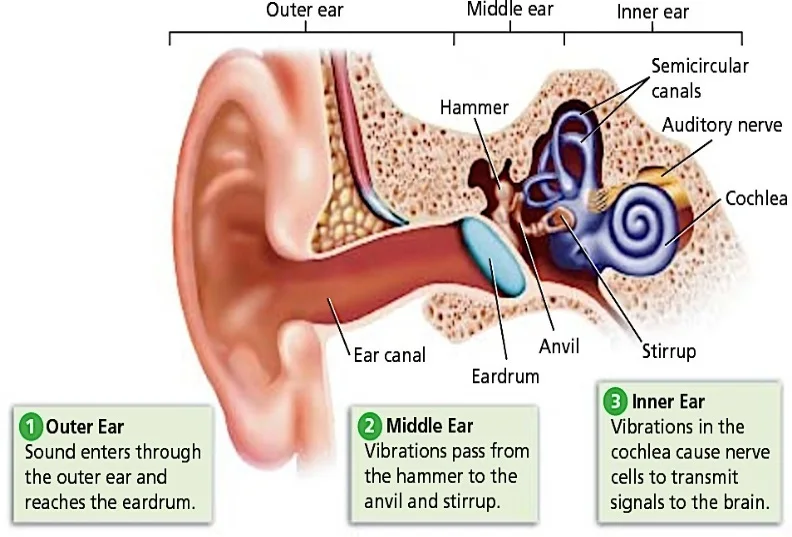
Your ears are paired organs, located on each side of your head.Human ear, organ of hearing and equilibrium that detects and analyzes sound by transduction and maintains the sense of balance.

What are the function of Ear?
The ears perform two sensory functions,
●Hearing and
●Maintenance of body balance (STATO-ACOUSTIC ORGAN)• Anatomically, the ear can be divided into three major sections called the outer ear, the middle ear and the inner ear.
Anatomy of Ear
THE OUTER EAR
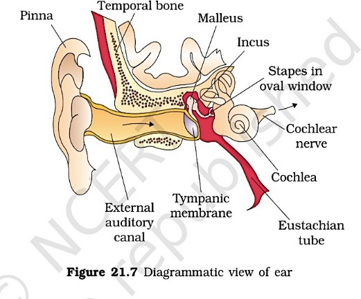
• The outer ear consists of the pinna and external auditory meatus (canal).
• The pinna collects the vibrations in the air which produce sound.
• The external auditory meatus leads inwards and extends up to the tympanic membrane ->the ear drum.
• There are very fine hairs and wax- secreting sebaceous glands in the skin of the pinna and the meatus.
• The tympanic membrane is composed of connective tissues covered with skin outside and with mucus membrane inside.

THE MIDDLE EAR
• It has three ossicles called malleus, incus and stapes (smallest bone of the body) crosses the tympanic cavity (cavity of middle ear) and are attached to one another in a chain-like fashion and increase the efficiency of transmission of sound waves to the inner ear
• The malleus is attached to the tympanic membrane and the stapes is attached to the oval window of the cochlea.
• Synovial hinge joint is present between mallus and incus whereas ball and socket joint are found between incus and stapes.
• Malleus is hammer shaped, incus is It is normally closed but open during swallowing and yawning.
• Fenestrae is a thin bony membrane between middle ear and inner ear and has two aperture, upper window is Fenestra ovalis and end of stapes fit on it. It is guarded by membrane.
• Fenestra rotundus is ventral window, it also connects middle ear to inner ear and it is locatedtowards Scala tympani.Outer ear,MIddle ear,Inner ear
THE INNER EAR
• The fluid-filled inner ear LABYRINTH.
• LABYRINTH consists of two parts, the bony labyrinth and the membranous labyrinths.
• The bony labyrinth is a series of channels.
• Membranous labyrinth presents inside bony labyrinth
• The cavity between bony and membranous labyrinth is called peri lymphatic space and filled with perilymph.
• The membranous labyrinth is filled with a fluid called ENDOLYMPH.
• The membranous labyrinth is consisting of two parts central sac like vestibule and cochlea.
• Vestibule has two chambers large utriculus and smaller sacculus.
• There is three semi-circular canal arises from utriculus and they are anterior semi-circular canal(upper), posterior semi-circular canal (Inferior) and horizontal / lateral semi-circular canal.
• Common part of anterior and posterior canal is crus commune and ampulla are terminal enlarged part of semi-circular canal.
• AMPULLA HAS SENSORY SPOT CALLED CRISTAE FOR EQUILIBRIUM.
• Crista is 3 in number; no otolith is found and has long auditory hair and it facilitates maintenance of dynamic equilibrium and angular acceleration.
• Sacculus is lower chamber of vestibule.
• Macula is group of sensory cells present in vestibules. In human 2 macula is present. It has otolith and short auditory hair and help in static equilibrium, linear acceleration that is tilting of head and rapid forward movement.
• The coiled portion of the labyrinth is called cochlea ; a short tube ductus reuniens which joins the cochlea with sacculus.
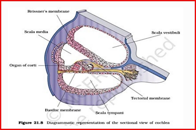
• Basilar membrane separates Scala media with Scala tympani.Sectional view of cochlea
• The membranes which make the cochlea are the Reisner’s and basilar membrane.

• Reisner membrane separates Scala media to scales vestibule
• These membranes divide the surounding perilymph filled bony labyrinth into an upper Scala vestibuli and a lower scala tympani.
• The space within cochlea called Scala media is filled with endolymph.
• At the base of the cochlea, the Scala vestibuli ends at the oval window, while the Scala tympani terminates at the round window which opens to the middle ear.The organ of corti
• It acts as auditory receptors.
• It is a structure located on the basilar membrane which contains hair cells.
• The hair cells are present in rows on the internal side of the organ of corti.
• The basal end of the hair cell is in close contact with the afferent nerve fibres.
• A large number of processes called stereo cilia are projected from the apical part of each hair cell.
• Above the rows of the hair cells is a thin elastic membrane called tectorial membrane.
Physiology & Hearing Mechanism:
• The vibrations produced in the ear drum are transmitted through the ear ossicles and oval window to the fluid-filled inner ear( cochlea), where they generate waves in the lymph.
• The waves in the lymph induce a ripple in the basilar membrane.
• These movements of the basilar membrane bend the hair cells, pressing them against the tectorial membrane. As a result, nerve impulses are generated in the associated afferent neurons.
• These impulses are transmitted by the afferent fibres via auditory nerves to the auditory cortex ofthe brain, where the impulses are analysed and the sound is recognised.
BODY POSTURE AND BALANCE OF THE BODY:
• The inner ear also contains a complex system called vestibular apparatus, located above thecochlea.
• The vestibular apparatus is composed of three semi-circular canals and the otolith organ consisting of the saccule and utricle.
• Each semi-circular canal lies in a different plane at right angles to each other.
• The membranous canals are suspended in the perilymph of the bony canals.
• The base of canals is swollen and is called ampulla, which contains a projecting ridge called CRISTA AMPULLARIS which has hair cells.
• The saccule and utricle contain a projecting ridge called macula.
• The crista and macula are the specific receptors of the vestibular apparatus WHICH ARE INFLUENCED BY GRAVITY and helps us in maintaining balance of the body and posture.
Ear Disorders
Outer Ear Disorders*
1. Otitis Externa (Swimmer’s Ear): Inflammation of the outer ear canal
2. Earwax Buildup: Excessive cerumen accumulation
3. Ear Canal Stenosis: Narrowing of the ear canal
4. Perichondritis: Inflammation of the outer ear cartilage
Middle Ear Disorders
1. Otitis Media (Middle Ear Infection): Bacterial or viral infection
2. Eustachian Tube Dysfunction: Blockage or dysfunction of the Eustachian tube
3. Tympanosclerosis: Scarring of the eardrum
4. Otosclerosis: Abnormal bone growth in the middle ear
Inner Ear Disorders
1. Benign Paroxysmal Positional Vertigo (BPPV): Balance disorder
2. Meniere’s Disease: Inner ear fluid imbalance
3. Labyrinthitis: Inner ear inflammation
4. Vestibular Migraine: Migraine-related balance disorder
Hearing Disorders
1. Conductive Hearing Loss: Middle ear problems
2. Sensorineural Hearing Loss: Inner ear or nerve damage3. Mixed Hearing Loss: Combination of conductive and sensorineural
4. Tinnitus: Ringing or other sounds in the ear
Balance Disorders
1. Vertigo: Spinning sensation
2. Dizziness: Lightheadedness or loss of balance
3. Balance Dysfunction: Difficulty with equilibrium
4. Motion Sickness: Nausea and balance disturbance
What are some symptoms of common ear conditions?
There are a number of symptoms that could indicate a problem with your ears. These warning signs include:
- Ear pain
- Ear infection.
- Clogged ears.
- Muffled hearing.
- Itchy ears.
- Nausea and vomiting.
- A feeling of fullness in your ears.
- Ear drainage.
TREATMENT
Treatment Options*
1. Medications
2. Surgery
3. Hearing aids
4. Cochlear implants
5. Vestibular rehabilitation therapy
6. Lifestyle modifications


Good 👍🏻
Thanks 😊
The human body has been created so amazingly.
Oh, interesting I have amazed too!