NEURAL CONTROL AND COORDINATION
NEURAL CONTROL AND COORDINATION
1. The neural system Function –> coordinates and integrates @ To maintain Homoeostasis therefore it is called neural control and coordination.
2. The neural system and the endocrine system jointly coordinate and integrate all the activities of the organs –> in a synchronised fashion.
3. The neural system –> An organised network of point-to-point connections for a quick coordination.
4. The neural system –> composed of highly specialised cells called NEURONS
5. Neurons can detect, receive and transmit different kinds of stimuli.
6. Ganglion cells /True nerve cell evolved for the first time in coelenterates.
7. Coelenterates nerve cell derive from interstitial cell and it is polar neurons.
8. In Platyhelminthes, ladder type of nervous system consists of cerebral ganglion/brain
9. Nematodes has central, peripheral and rectal nervous system and rectal nervous system develop more in male.
10. In annelids, nervous system consists of central N. S., peripheral N.S. and sympathetic nervous system. CNS is made up of nerve ring and ventral nerve cord.
11. In arthropods all three nervous systems (central N. S., peripheral N.S. and sympathetic nervous system) are present; Sympathetic nervous system (in cockroach) also called somatogastric nervous system.
12. In gastropods (Mollusca) like pila consist of paired ganglion, commissures and connective.
13. In some Mollusca, which have sluggish and sedentary life, nervous system is reduced like in pelecypod (unio).
14. In cephalopods –> Nervous system highly evolved. @ Octopus
15. In Echinodermates, it is simple and primitive and consist of nerve fibres and few ganglion cells, all are confined to body wall except the visceral nerve plexus situated at the gut wall.
16. Hemichordates have primitive type of nervous system and it is consisting of ventral and dorsal nerve cord.
17. In chordates, it is well developed and formed from ectoderm. It has central nervous system, peripheral nervous system and autonomic nervous system.
HUMAN NEURAL SYSTEM.
18. The human neural system is divided into TWO PARTS:
● The Central Nervous System
●The Peripheral Nervous System,
19. The central neural systems (CNS): The CNS includes the brain and the spinal cord and is the site of information processing and control.
20. Nervous system begins developing early in third week of embryonic development.
21. In nervous system only MICROGLIAL CELL develop from mesoderm which are specialised population of macrophages that are found in CNS and remove damaged neurons and infection. THEY ARE NECESSARY FOR MAINTENANCE OF CNS.
22. Germinal layer of neural tube form ventricles of brain and lining of neural canal called EPENDYMA (ciliated columnar epithelium).
●Mantle layer of neural tube consists of embryonic neuron” nematoblast” and form grey matter.
●Marginal layer of neural tube form white matter.
●Enlarged anterior part of neural tube form embryonic brain called ENCEPHALON.

●Encephalon by differential growth and two constriction, it is divided in to three primary vesicles called forebrain, midbrain and hind brain which develop into thee major division of adult brain called —
(1) prosencephalon/forebrain which has two division anterior telencephalon and posterior diencephalon,
(2) Mesencephalon /midbrain and
(3) Rhombencephalon/hindbrain which has two division anterior
metencephalon and posterior myelencephalon.
●In all vertebrates CNS is dorsal, hollow and non-ganglionated.
23. The brain is the central information processing organ of our body, and acts as the ‘command and control system’.
24. It controls the
● Voluntary movements,
● Balance of the body,
●Functioning of vital involuntary organs (e.g., lungs, heart, kidneys, etc.),
● Thermoregulation,
● Hunger and thirst,
● Circadian (24-hour) rhythms of our body,
● Activities of several endocrine glands and
●Human behaviour.
25. It is also the site for processing of vision, hearing, speech, memory, intelligence, emotions and thoughts.
26. The human brain is well protected by the skull.
27. Inside the skull, the brain is covered by cranial meninges consisting of:
● DURA MATER -> Outer most layer; It is tough, non-vascular and made up of fibrous connective tissue.
●ARACHNOID -> Very thin middle layer ; reticular connective tissue.
●PIAMATER -> an inner layer (which is in contact with the brain tissue) ; consist of loose aeriolar tissue and vascular and nutritive in nature.
Falx caribri is extension of duramatter present between cerebral hemisphere ; Tentorium is duramater extension present between cerebrum and cerebellum.
28. The brain can be divided into three major parts:
■Forebrain,
■ Midbrain, and
■ Hindbrain
29. Forebrain:
●The forebrain consists of cerebrum, thalamus and hypothalamus.
30. Cerebrum – It forms the major part of the human brain.
31. Cerebrum is divided into 5 parts called frontal, parietal, occipital, temporal, and insula and insula is hidden sylvian fissure.
32. The median fissure divides cerebrum into right and left cerebral hemisphere.
33. Rhinal fissure separates olfactory lobes from cerebral hemisphere.,
34. Cerebral hemisphere has fluid filled cavity called paracoel or lateral ventricle.
35. Corpous callosum is a tract of nerve fibre of white matter which connect the two halves of cerebrum.
36. Corpus callosum is characteristics of EUTHERIANS MAMMALS.
37. Degeneration or improper development of corpus callosum is result into SCHIZOPHRENIA.
38. The layer of cells which covers the cerebral hemisphere is called cerebral cortex/pallium and is thrown into prominent folds.
39. The cerebral cortex is referred to as the grey matter due to its greyish appearance.
40. The neuron cell bodies are concentrated here giving the grey colour.
41. The cerebral cortex contains –>
● Motor areas, sensory areas and large regions that are neither clearly sensory nor motor in function.
●These regions called as the association areas are responsible for complex functions like inter-sensory associations, memory and communication.
42. Cerebral medulla that is deeper part of hemisphere is made up of white matter.
43. IF CEREBRUM IS REMOVED –> Animal become simple reflex organism.
44. Diencephalon cavity is called DIOCOEL or 3rd RD VENTRICLE.
● The thin roof of this cavity is EPITHALAMUS,the thick right and left side is called THALAMUS and floor of this cavity is HYPOTHALAMUS.
45. The cerebrum wraps around a structure called thalamus, which is a major coordinating centre for sensory and motor signaling. It consist 80% of diencephalon.

46. HYPOTHALAMUS:
• It lies at the base of the thalamus.
• The hypothalamus contains a number of centres which control body temperature, urge for eating and drinking.
• It also contains several groups of neurosecretory cells (pituitary gland/ hypophysis) , which secrete
hormones called hypothalamic hormones.
• In front of hypothalamus, optic chiasma (responsible for binocular vision) found and it, one pair of small rounded mamillary body/corpora mammillarias found.
• The inner parts of cerebral hemispheres and a group of associated deep structures like amygdala,
hippocampus, etc., form a complex structure called the LIMBIC LOBE OR LIMBIC SYSTEM.
• Limbic system and the hypothalamus combindly involved
o In the regulation of sexual behaviour,
o Expression of emotional reactions (e.g., excitement, pleasure, rage and fear), and motivation.
• Pineal gland located at centre of brain, innervated by sympathetic nerve produce a hormone called melatonin which is anti FSH and LH . It inhibits reproductive function but after puberty, melatonin secretion is decreased.
• FUNCTION OF FORE BRAIN
o Centre of smell, It confirms, recognises and evaluates shape,colour,sound ,taste and smell for sensory cell in relation with object,
o BROCA’S AREA –-> Translate thought into speech also associated with language area. Damage in this area result into aphasia (inability to speak), deafness and blindness.
o Somaesthetic area is for perception of pain, touch and temperature.
o Premotor area in frontal lobe is centre of involuntary movement of muscle and ANS.
o Cerebrum is centre of intelligence, emotion, memory ,will power, weeping, laughing, reasoning, voluntary control, micturation and defecation
o Diencephalon is centre of carbohydrate, fat metabolism.
o HYPOTHALAMUS It is centre of hunger, thirst, sweating, sleep, fatigue, anger, pleasure, love hate and satisfaction and also control autonomic nervous system.

MIDBRAIN
• The midbrain is located between the thalamus/hypothalamus of the forebrain and pons of the hindbrain.
• Posterior part of mid brain is tectum.
• It contains both grey and white matter.
• The dorsal portion of the
midbrain consists mainly of four round swellings (lobes) called CORPORA QUADRIGEMINA.
• A canal called the CEREBRAL AQUEDUCT passes through the midbrain.
• MIDBRAIN is related with vision and auditory function.
• The corpora quadrigemina are reflex centers involving vision and hearing the reflex centres of eye movement and auditory responses.
• Midbrain basically connects the forebrain and the hind brain.

• HIND BRAIN
o It consists of
●CEREBELLUM,
●MEDULLA OBLONGATA
●PONS VAROLII. CEREBELLUM
• hind brain Also called sandwiched brain
• second largest portion after cerebrum.
• It is butterfly in shape.
• It has vermis, the central constricted area,
lateral wings called cerebellar hemisphere which govern skeletal muscles movement, and the flocculo nodular lobe which is related with SENSE OF EQUILIBRIUM.
• When a person drinks alchohol, cerebellum involve in loss of control. MEDULLA OBLONGATA
• Its cavity is metacoel / IVth ventricle which is continuous with central canal of spinal cord.
• It has a pair of LATERAL FORAMEN OF LUSHAKA and a median FORAMEN OF MAGENDIE through these aperture, CSF of cavity of brain and fluid of meninges come in contact with each other.
• In medulla oblongata, most of the sensory and motor nerve fibre cross each other therefore left cerebral hemisphere controls right side of body and vice versa.
• Medulla contain 5 pair of cranial nerve from VIII to XII.
• It controls heartbeat, respiration, digestion, BP, swallowing of food, urination, defecation, vomiting, coughing, hiccupping, sneezing, vasodilator and vasoconstrictor etc
PONS VAROLII
• Oval mass of white matter
• Located above the medulla oblongata.
• It has pneumotaxic & apneustic area.
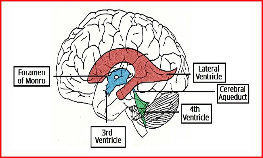
• The pneumotaxic center controls both the rate and the pattern of breathing. VENTRICLES OF BRAIN:
o RHINOCOEL olfactory lobe.
o Lateral ventricle /paracoel/ I and II ventricle cavity of cerebrum.
o FORAMEN OF MONRO lateral ventricles connect with IIIrd ventricle through this foramen.
o Diocoel is IIIrd ventricle.
o AQUIDUCT OF SYLVIUS is narrow cavity between III and IV th ventricle.
o OPTOCOEL is 4th ventricle of optic lobe.
• CEREBELLUM is solid.
• Midbrain and hindbrain form the brain stem.
• The hindbrain comprises pons, cerebellum and medulla (also called the medulla oblongata).
• PONS CONSISTS OF FIBRE TRACTS THAT INTERCONNECT DIFFERENT REGIONS OF THE BRAIN.
• Cerebellum has very convoluted surface in order to provide the additional space for many more neurons.
• The medulla of the brain is connected to the spinal cord. It has centres which control respiration,
cardiovascular reflexes and gastric secretions.

The peripheral neural system (PNS)
47. The PNS comprises of all the nerves of the body associated with the CNS (brain and spinal cord).
48. The nerve fibres of the PNS are of two types:
(a) Afferent fibres/sensory nerve: They transmit impulses from tissues or organs to the CNS.
(b) Efferent fibres/motor nerve: They transmit regulatory impulses from the CNS to the concerned peripheral tissues or organs.
49. The PNS is divided into two divisions called
(1) Somatic neural system: The somatic neural system relays impulses from the CNS to skeletal muscles.
(2) Autonomic neural system: The autonomic neural system transmits impulses from the CNS to the involuntary organs and smooth muscles of the body.
The autonomic neural system is further classified into sympathetic neural system and parasympathetic neural system.
In SYMPATHETIC AUTONOMIC NEURAL SYSTEM, postgangliotic nerve secrete SYMPATHETIN (NOR- EPINEPHRINE) AND ACETYLCHOLINE both, expenditure of energy take place and IT IS ACTIVE IN STRESS, PAIN, FEAR AND ANGER CONDITION. It increase defence system of body in adverse condition while PARASYMPATHETIC AUTONOMIC NEURAL SYSTEM secretes acetylcholine and provide relaxation, comfort, pleasure at the time of rest. It is related with restoration and conservation of energy.
NEURON

Act AS STRUCTURAL AND FUNCTIONAL UNIT OF NEURAL SYSTEM
• A neuron is a microscopic structure composed of three
major parts namely, CELL BODY
• The cell body contains cytoplasm with typical cell
organelles and certain granular bodies called Nissl’s granules.
@These granules are derived from Rough ER DENDRITES
• Branched Short fibres which branch project out of the cell
body also contain Nissl’s granules are called dendrites. These fibres transmit impulses towards the cell body.
AXON
• A long fibre with branched distil end.
• Each branch of distil end terminates as a bulb-like structure called SYNAPTIC KNOB which possess SYNAPTIC VESICLES containing chemicals called NEUROTRANSMITTERS.
• The axons transmit nerve impulses away from the cell body to a synapse or to a neuro-muscular junction.
50. Based on the number of axon and dendrites, the neurons are divided into three types-:
(i) Multipolar: It is with one axon and two or more dendrites and found in the cerebral cortex.
(ii) Bipolar: It is with one axon and one dendrite and found in the retina of eye.
(iii) Unipolar: cell body with one axon only; found usually in the embryonic stage.
51. There are two types of axons, namely, myelinated and nonmyelinated.
MYELINATED NERVE
●Enveloped with Schwann cells which form a myelin sheath around the axon.
● The gaps between two adjacent myelin sheaths are called NODES OF RANVIER.
Myelinated nerve fibres are found in spinal and cranial nerves.
NON-MYELINATED NERVE FIBRE
● It is enclosed by a Schwann cell that does not form a myelin sheath around the axon and is commonly found in autonomous and the somatic neural systems.
GENERATION AND CONDUCTION OF NERVE IMPULSE:

52. NEURONS ARE EXCITABLE -> Due to presence of different ion channels in neural membrane neurons @ these channels are selective permeable.
53. NEURON IN RESTING STAGE Have resting membrane potential (-70 mv) ; POLARISED STATE @ because the axonal membrane is comparatively more permeable to potassium ions (K+) and nearly impermeable to sodium ions (Na+). Similarly, the membrane is impermeable to negatively charged proteins present in the axoplasm therefore the axoplasm(cytoplasm) inside the axon contains high concentration of K+ and negatively charged proteins and low concentration of Na+.
54. The fluid outside the axon contains a low concentration of K+, a high concentration of Na+ and thus form a concentration gradient.
55. These ionic gradients across the resting membrane are maintained by the active transport of ions by the sodium-potassium pump which transports 3 Na+ outwards for 2 K+ into the cell.
56. Because of these concentration gradient, the outer surface of the axonal membrane possesses a positive charge while its inner surface becomes negatively charged and therefore is polarised.
57. The electrical potential difference across the resting plasma membrane is called as the RESTING POTENTIAL.
GENERATION OF NERVE IMPULSE
58. When a stimulus is applied at a site on the polarised membrane, the membrane at that site becomes freely permeable to Na+. This leads to a rapid influx of Na+ followed by the reversal of the polarity at that site, i.e., the outer surface of the membrane becomes negatively charged and the inner side becomes positively charged.
59. The polarity of the membrane at that site is thus reversed and hence depolarised.
60. The electrical potential difference across the plasma membrane at that site is called the ACTION POTENTIAL (a nerve impulse).
61. At sites immediately ahead, the axon membrane has a positive charge on the outer surface and a negative charge on its inner surface(polarised state). As a result, a current flow on the inner surface from that site to site ahead.
62. On the outer surface of membrane, current flows from site ahead to previous site to complete the circuit of current flow.
63. Hence, the polarity at the site is reversed, and an action potential is generated at site ahead. Thus, the impulse (action potential) generated at previous site arrives at site ahead.
64. The sequence is repeated along the length of the axon and consequently the impulse is conducted.
65. The rise in the stimulus-induced permeability to Na+ is extremely short-lived.
66. It is quickly followed by a rise in permeability to K+.
67. Within a fraction of a second, K+ diffuses outside, the membrane and restores the resting potential of the membrane at the site of excitation and the fibre becomes once more responsive to further stimulation.
Transmission of Impulses from one neuron to next neuron
How synapse work
Subscribe youtube channel

68. A nerve impulse is transmitted from one neuron to another through junctions called SYNAPSES.
69. A synapse is formed by the membranes of a pre-synaptic neuron and a post-synaptic neuron(next neuron), which may or may not be separated by a gap called synaptic cleft.
70. There are two types of synapses:
• Electrical synapses.
• Chemical synapses.
ELECTRICAL SYNAPSES
●Electrical synapses are rare in our system,
●Impulse transmission across an electrical synapse is always faster than that across a chemical synapse.
● Transmission of an impulse across electrical synapses is very similar to impulse conduction along a single axon as the membranes of pre- and post-synaptic neurons are in very close proximity (synaptical cleft is absent) so the electrical current can flow directly from one neuron into the other across these synapses.
CHEMICAL SYNAPSES
●the entry of ions which can generate a new action potential Here, the membranes of the pre- and post-synaptic neurons are separated by a fluid-filled space called SYNAPTIC CLEFT.
● Chemicals called neurotransmitters are involved in the transmission of impulses at these synapses.
●The axon terminals contain vesicles filled with these neurotransmitters.
● When an impulse (action potential) arrives at the axon terminal, it stimulates the movement of the synaptic vesicles towards the membrane where they fuse with the plasma membrane and release their neurotransmitters in the synaptic cleft.
●The released neurotransmitters bind to their specific receptors, present on the post-synaptic membrane.
● This binding opens ion channels allowing in the post-synaptic neuron.
● The new potential developed may be either excitatory or inhibitory.
REFLEX ACTION AND REFLEX ARC

• Involuntary response to a peripheral nervous stimulation of reflex action.
• It is without conscious effort or thought and requires the involvement of a part of the CNS @SPINAL CORD
• THE REFLEX PATHWAY COMPRISES
●One afferent neuron (receptor)
●One efferent (effector or excitor) Neuron appropriately arranged in a series.
• The afferent neuron receives signal from a sensory organ and transmits the impulse via a dorsal nerve root into the CNS (at the level of spinal cord).
• The efferent neuron then carries signals from CNS to the effector. The stimulus and response thus form a REFLEX ARC.
SENSORY RECEPTION AND PROCESSING:
• Information regarding changes in the environment is received by the CNS through the sensory organs which are processed and analysed.
• Signals are then sent for necessary adjustments.
Eye

●One paired eyes are located in sockets of the skull called orbits.
●The wall of the eye ball is composed of three layers .
●SCLERA: External layer which is composed of a dense connective tissue. IT IS WHITE PART OF EYE.
CORNEA: The anterior portion of sclera is called the cornea.
CHOROID:
●The middle layer, choroid, contains many blood vessels and looks bluish in colour.
●The choroid layer is thin over the posterior two-thirds of the eye ball, but it becomes thick in the anterior part to form the ciliary body.
●The ciliary body itself continues forward to form a pigmented and opaque structure called the iris which is the visible coloured portion of the eye (give eye colour).
●Ciliary body help in accommodation by altering the focus of eye.
IRIS AND CILLIARY BODY IS PART OF CHOROID LAYER. LENS:
o The eye ball contains a transparent crystalline lens which is held in place by suspensory ligaments (Zonula of Zinn) attached to the ciliary body.
LENS DIVIDE THE EYE BALL INTO OUTER AQUEOUS AND INNER VITREOUS CHAMBER.
PUPIL: In front of the lens, the aperture surrounded by the iris is called the pupil. The diameter of the pupil is regulated by the muscle fibres of iris.
RETINA: The inner layer is the retina and it contains three layers of cells – from inside to outside –
●Ganglion Cells,
●Bipolar Cells &
●Photoreceptor Cells:
• There are two types of photoreceptor cells -> rods and cones.
• These cells contain the light-sensitive proteins called the photopigments.
• The daylight (photopic) vision and colour vision are functions of cones.
• Photoreceptor cells are not present in that region and hence it is called the blind spot.
• The twilight (scotopic) vision is the function of the rods.
• The rods contain a purplish-red protein called the rhodopsin or visual purple, which contains a derivative of Vitamin A.
• In the human eye, there are three types of cones which possess their own characteristic photopigments that respond to red, green and blue lights.
• The sensations of different colours are produced by various combinations of these cones and their
photopigments.
• When these cones are stimulated equally, a sensation of white light is produced.
• The optic nerves leave the eye and the retinal blood vessels enter it at a point medial to and slightly above the posterior pole of the eye ball.
• At the posterior pole of the eye lateral to the blind spot, there is a yellowish pigmented spot called MACULA LUTEA with a central pit called the FOVEA.
• The fovea is a thinned-out portion of the retina where only the cones are densely packed. It is the
point where the visual acuity (resolution) is the greatest.
• Aqueous chamber and humor: The space between the cornea and the lens is called the AQUEOUS CHAMBER and contains a thin watery fluid called aqueous humor.
• Vitreous chamber: The space between the lens and the retina is called the VITREOUS CHAMBER and is filled with a transparent gel called vitreous humor.
MECHANISM OF VISION
• The light rays in visible wavelength focussed on the retina through the cornea and lens generate potentials (impulses) in rods and cones.
• The photosensitive compounds (photopigments) in the human eyes is composed of opsin (a
protein) and retinal which is an aldehyde of vitamin A.
• Light induces dissociation of the retinal from opsin resulting in changes in the structure of the opsin. This causes membrane permeability changes.
• As a result, potential differences are generated in the photoreceptor cells.
• This produces a signal that generates action potentials in the ganglion cells through the bipolar cells.
• These action potentials (impulses) are transmitted by the OPTIC NERVES to the VISUAL CORTEX
AREA OF THE BRAIN, where the neural impulses are analysed and the image formed on the retina is recognised based on earlier memory and experience.
• Eye glands
o Meibomian gland -> Lubrication of eye.
o Lacrimal gland -> tear production; tear is slightly saline and it moist the eye.
o Glands of zeis -> lubricate hair follicle in eye lid
o Glands of moll -> It open into follicles of eye lashes.
• Human has day and night vision both as it has rods and cones both.
EYE DEFECTS
• Myopia/ near -sightedness In this eye ball become longer and image is formed before retina and can be removed by use of concave lens.
• Hypermetropia/ far-sightedness In this eye ball become short so image form behind the retina and can be removed by use of convex lens.
• ASTIGMATISM is curvature of cornea become irregular and image is not form clearly and this defect
can be cured by use of cylindrical lens.
• CATARACT is due to defective protein metabolism. Lens or cornea become opaque and operation is needed to cure this.
• XEROPHTHALMIA due to deficiency of vitamin A, cornea /conjunctiva become keratinised which may lead to blindness.
• GLAUCOMA due to increase in intraocular pressure in aqueous chamber and surgery is needed at early stage due to blockage of Schlemm’s canal.
• STRABISMUS is associated with squintness. In which eye is remained in somewhat bended position.
• TRACHOMA redness of eye and more secretion of watery fluid and it due to infection of bacteria
chlamydia trachomatis.
REMEMBER:
4 rectus and 2 oblique muscles are associated with eye ball movement.

THE EAR
• The ears perform two sensory functions,
●Hearing and
●Maintenance of body balance (STATO-ACOUSTIC ORGAN)
• Anatomically, the ear can be divided into three major sections called the outer ear, the middle ear and the inner ear.
THE OUTER EAR
• The outer ear consists of the pinna and external auditory meatus (canal).
• The pinna collects the vibrations in the air which produce sound.
• The external auditory meatus leads inwards and extends up to the tympanic membrane ->the ear drum.
• There are very fine hairs and wax- secreting sebaceous glands in the skin of the pinna and the meatus.
• The tympanic membrane is composed of connective tissues covered with skin outside and with mucus membrane inside.
THE MIDDLE EAR
• It has three ossicles called malleus, incus and stapes (smallest bone of the body) crosses the tympanic cavity (cavity of middle ear) and are attached to one another in a chain-like fashion and increase the efficiency of transmission of sound waves to the inner ear.
• The malleus is attached to the tympanic membrane and the stapes is attached to the oval window of the cochlea.
• Synovial hinge joint is present between mallus and incus whereas ball and socket joint are found
between incus and stapes.
• Malleus is hammer shaped, incus is It is normally closed but open during swallowing and yawning.
• Fenestrae is a thin bony membrane between middle ear and inner ear and has two aperture, upper window is Fenestra ovalis and end of stapes fit on it. It is guarded by membrane.
• Fenestra rotundus is ventral window, it also connects middle ear to inner ear and it is located
towards Scala tympani.

THE INNER EAR
• The fluid-filled inner ear LABYRINTH.
• LABYRINTH consists of two parts, the bony labyrinth and the membranous labyrinths.
• The bony labyrinth is a series of channels.
• Membranous labyrinth presents inside bony labyrinth
• The cavity between bony and membranous labyrinth is called peri lymphatic space and filled with perilymph.
• The membranous labyrinth is filled with a fluid called ENDOLYMPH.
• The membranous labyrinth is consisting of two parts central sac like vestibule and cochlea.
• Vestibule has two chambers large utriculus and smaller sacculus.
• There is three semi-circular canal arises from utriculus and they are anterior semi-circular canal(upper), posterior semi-circular canal (Inferior) and horizontal / lateral semi-circular canal.
• Common part of anterior and posterior canal is crus commune and ampulla are terminal enlarged part of semi-circular canal.
• AMPULLA HAS SENSORY SPOT CALLED CRISTAE FOR EQUILIBRIUM.
• Crista is 3 in number; no otolith is found and has long auditory hair and it facilitates maintenance of dynamic equilibrium and angular acceleration.
• Sacculus is lower chamber of vestibule.
• Macula is group of sensory cells present in vestibules. In human 2 macula is present. It has otolith and short auditory hair and help in static equilibrium, linear acceleration that is tilting of head and rapid forward movement.
• The coiled portion of the labyrinth is called cochlea ; a short tube ductus reuniens which joins the cochlea with sacculus.
• Basilar membrane separates Scala media with Scala tympani.

• The membranes which make the cochlea are the Reisner’s and basilar membrane.
• Reisner membrane separates Scala media to scales vestibule
• These membranes divide the surounding perilymph filled bony labyrinth into an upper Scala vestibuli and a lower scala tympani.
• The space within cochlea called Scala media is filled with endolymph.
• At the base of the cochlea, the Scala vestibuli ends at the oval window, while the Scala tympani terminates at the round window which opens to the middle ear.
The organ of corti
• It acts as auditory receptors.
• It is a structure located on the basilar membrane which contains hair cells.
• The hair cells are present in rows on the internal side of the organ of corti.
• The basal end of the hair cell is in close contact with the afferent nerve fibres.
• A large number of processes called stereo cilia are projected from the apical part of each hair cell.
• Above the rows of the hair cells is a thin elastic membrane called tectorial membrane.
HEARING MECHENISM:
• The vibrations produced in the ear drum are transmitted through the ear ossicles and oval window to the fluid-filled inner ear( cochlea), where they generate waves in the lymph.
• The waves in the lymph induce a ripple in the basilar membrane.
• These movements of the basilar membrane bend the hair cells, pressing them against the tectorial membrane. As a result, nerve impulses are generated in the associated afferent neurons.
• These impulses are transmitted by the afferent fibres via auditory nerves to the auditory cortex of
the brain, where the impulses are analysed and the sound is recognised.
BODY POSTURE AND BALANCE OF THE BODY:
• The inner ear also contains a complex system called vestibular apparatus, located above the
cochlea.
• The vestibular apparatus is composed of three semi-circular canals and the otolith organ consisting of the saccule and utricle.
• Each semi-circular canal lies in a different plane at right angles to each other.
• The membranous canals are suspended in the perilymph of the bony canals.
• The base of canals is swollen and is called ampulla, which contains a projecting ridge called CRISTA AMPULLARIS which has hair cells.
• The saccule and utricle contain a projecting ridge called macula.
• The crista and macula are the specific receptors of the vestibular apparatus WHICH ARE INFLUENCED BY GRAVITY and helps us in maintaining balance of the body and posture.
SOME IMPORTANT FACTS
• Proprioceptorss is located in skeletal muscles , joints and tendons and through these receptors we know the position of our arms and legs without having a look into it.
• Tangoreceptor is sensitive to touch pressure.
• Thigmoreceptor and tectoreceptor is related with touch sensation.
• 12 pairs of cranial nerve are present in reptiles, birds and mammals.
• The smallest cranial nerve in human is trochlear but, in all animals, it is abducens.
• The largest cranial nerve in human is trigeminal but in all animals it is vagus nerve.
• Cerebrospinal fluid/CSF is secreted by the choroid plexus by filtration of blood.
• Hydrocephalus is enlargement of head by abnormal accumulation of CSF resulting in pain, vomiting and stiffness of neck.
• Increased CSF may result into Meningitis. It may occur due to infection and inflammation of
meninges or injury of meninges. Infection can be viral or bacterial.
• Lumber puncture is done to drain excess of CSF.
Comment your review

One thought on “NEURAL CONTROL AND COORDINATION”
You must log in to post a comment.