🧬 Human Reproduction: Male & Female Reproductive System Explained (Easy Notes + Diagrams)
Introduction
human reproduction
NOTE – Please note there may be error in diagram labelling
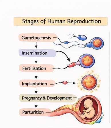
Human beings reproduce sexually and are viviparous, meaning they give birth to live young ones. Reproduction involves:
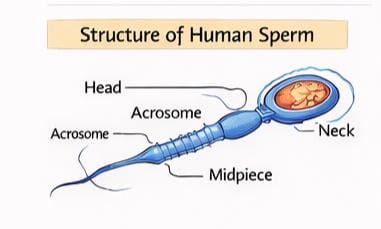
- Gametogenesis – formation of male (sperm) & female (ovum) gametes
- Insemination – transfer of sperm into female tract
- Fertilisation – fusion of gametes
- Implantation – attachment of blastocyst to uterus
- Pregnancy & Gestation – development of embryo
- Parturition – childbirth
- Lactation – milk secretion post-birth
Let’s understand each system and process in detail.
1. Male Reproductive System
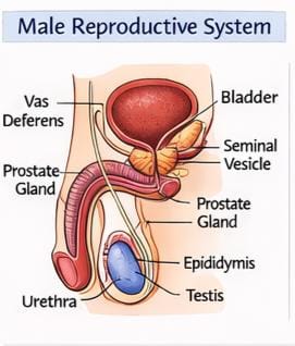
The male reproductive organs lie in the pelvic region and consist of:
1.1 Testes
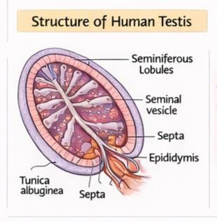
- Located inside the scrotum (2–2.5°C cooler for spermatogenesis).
- Each testis: 4–5 cm long, 2–3 cm wide.
- Contain 250 testicular lobules.
- Each lobule has 1–3 seminiferous tubules where sperms are produced.
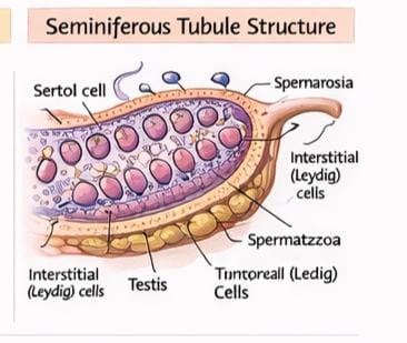
1.2 Cells in Seminiferous Tubules
- Spermatogonia – male germ cells → form sperms.
- Sertoli cells – nourish developing sperms.
- Leydig cells (in interstitial space) – secrete androgens (testosterone).
1.3 Male Accessory Ducts
- Rete testis
- Vasa efferentia
- Epididymis
- Vas deferens → forms ejaculatory duct → opens into urethra
1.4 Male Accessory Glands
- Seminal vesicles
- Prostate gland
- Bulbourethral (Cowper’s) glands
Their secretions form seminal plasma, rich in:
- Fructose
- Calcium
- Enzymes
1.5 Penis
- Male external genitalia
- Glans penis covered by foreskin
- Special erectile tissue enables penetration
2. Female Reproductive System
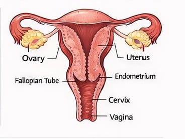
Consists of:
- Ovaries
- Oviducts (fallopian tubes)
- Uterus
- Cervix
- Vagina
- External genitalia
- Mammary glands
2.1 Ovaries
- Primary female sex organs
- Produce ovum + estrogen & progesterone
- Contain ovarian follicles in various stages (primary → secondary → Graafian follicle)
2.2 Fallopian Tubes
- 10–12 cm long
- Divided into infundibulum, ampulla, isthmus
- Fimbriae collect egg during ovulation
- Fertilisation occurs in ampulla
2.3 Uterus
- Inverted pear-shaped
- Three layers:
- Perimetrium
- Myometrium (strong muscle for childbirth)
- Endometrium (sheds during menstruation)
2.4 Vagina & External Genitalia
Includes:
- Mons pubis
- Labia majora
- Labia minora
- Hymen
- Clitoris
2.5 Mammary Glands
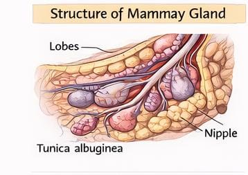
- 15–20 lobes with alveoli
- Store and secrete milk after childbirth
3. Gametogenesis
3.1 Spermatogenesis
Occurs in seminiferous tubules.
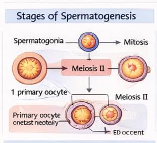
Steps:
- Spermatogonia → divide mitotically
- Form primary spermatocytes
- Meiosis I → secondary spermatocytes
- Meiosis II → spermatids
- Spermiogenesis → spermatids become sperm
- Spermiation → release of sperm into lumen
Key hormones:
- GnRH → stimulates LH & FSH
- LH → stimulates Leydig cells (androgens)
- FSH → helps Sertoli cells promote spermiogenesis
Normal ejaculation: 200–300 million sperms
3.2 Oogenesis
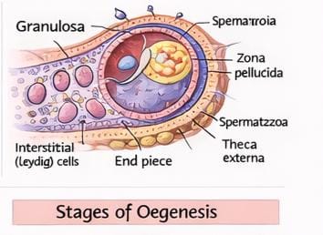
Begins before birth.
Steps:
- Oogonia → primary oocytes (arrested in prophase-I)
- At puberty: primary oocyte → secondary oocyte + polar body
- During ovulation → secondary oocyte released
- Meiosis completes only after fertilisation
4. Menstrual Cycle (28 Days)
Phases:
- Menstrual Phase (Day 1–5)
- Endometrial shedding
- Occurs if no fertilisation
- Follicular Phase (Day 6–13)
- Follicle grows
- Endometrium regenerates
- Estrogen increases
- FSH & LH rise
- Ovulation (Day 14)
- LH surge → Graafian follicle ruptures → ovulation
- Luteal Phase (Day 15–28)
- Corpus luteum forms → secretes progesterone
- Maintains endometrium
- If no pregnancy → corpus luteum degenerates → menstruation
5. Fertilisation & Implantation
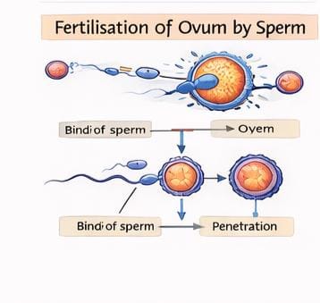
Steps:
- During coitus → sperm enters vagina
- Sperm travels to ampulla of fallopian tube
- Sperm penetrates zona pellucida
- Fusion of gametes → zygote (46 chromosomes)
- Cleavage → 2, 4, 8, 16-cell morula
- Morula → blastocyst
- Implantation occurs into endometrium → pregnancy starts
Sex determination happens at fertilisation:
- XX → female
- XY → male
Father determines sex of baby.
6. Pregnancy & Embryonic Development
Placenta
Formed by:
- Chorionic villi
- Maternal uterine tissue
Functions:
- Nutrient & oxygen supply
- Removal of waste
- Hormone production: hCG, hPL, progesterone, estrogen, relaxin
Development Timeline
- 1 month → heart formed
- 2 months → limbs + digits
- 12 weeks → major organs developed
- 5 months → movement + hair
- 24 weeks → eyelids separate, eyelashes
- 9 months → fully developed baby
7. Parturition (Childbirth)
Triggered by:
- Mature fetus
- Placental signals
- Oxytocin → strong uterine contractions → delivery
- After baby → placenta expelled
8. Lactation
- Mammary glands produce milk
- First milk = colostrum
- Rich in antibodies
- Essential for newborn immunity
Conclusion
Human reproduction is a complex but beautifully coordinated sequence of events involving gamete formation, fertilisation, implantation, pregnancy, childbirth, and nourishment. Understanding these processes is essential for biology students and anyone interested in human physiology.
read also Cell cycle and cell division
follow facebook.com/sciencegajab/
NOTE – Please note there may be error in diagram labelling do not trust blindly on labelling