
Your muscular heart, the main organ in your cardiovascular system, is vital for life.
Anatomy of heart:
Location: The heart is located in the center of the chest, near the lungs.
Chambers: It has four chambers: two upper chambers called atria (right and left) and two lower chambers called ventricles (right and left).
Valves: Heart valves are located at the openings of the chambers and ensure blood flows in the correct direction.
Muscular Tissue: The heart – made up of muscle and other heart tissue.
Pericardium: The heart – surrounded by a tough membrane called the pericardium.
What is the function of the heart?
Your heart’s main function is to move blood throughout your body. Blood brings oxygen and nutrients to your cells. It also takes away carbon dioxide and other waste so other organs can dispose of them.
How to Read Electrocardiograph (ECG)?
Electro-cardiograph is a device used to obtain an electrocardiogram (ECG).
To obtained a standard ECG, patient is connected to the machinewith three electrical leads, two in wreast and one in left ankle which monitor the heart activity.
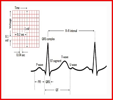
ECG is a graphical representation of the electrical activity of the heart during a cardiac cycle.
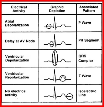
Each peak in the ECG is identified with a letter from P to T that corresponds to a specific electrical activity of the heart.
The P-wave represents the electrical excitation (or depolarisation) of the atria, which leads to the contraction/systole of both the atria.
The QRS complex represents the depolarisation of the ventricles, which initiates the ventricular contraction.
The contraction starts shortly after Q and marks the beginning of the systole.
The T-wave represents the return of the ventricles from excited to normal state (repolarisation).
The end of the T-wave marks the end of systole.
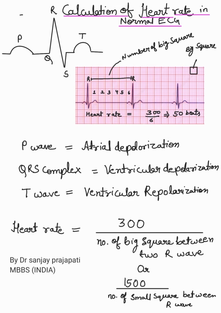
By counting the number of QRS complexes that occur in a given time period, one can determine the heart beat rate of an individual.
Any deviation from normal shape indicates a possible abnormality or disease. Hence, it is of a great clinical significance.
Any abnormality in above wave indicates the disorder of corresponding part of heart structure to the wave.
Calculations of heart rate in normal ECG
also click for better understanding of heart
https://www.instagram.com/sciencegajab


She does an EKG differ from an EEG – echocardiogram?
Ekg also known as Ecg records electrical activity of heart
Whereas EEG (ELECTROENCEPHALOGRAM)RECORDS electrical activity of your brain 🧠 .
https://forbesscotland.com/simonetta-lein-advocates-for-positive-change-in-entertainment-with-inclusive-message/
Visit it also